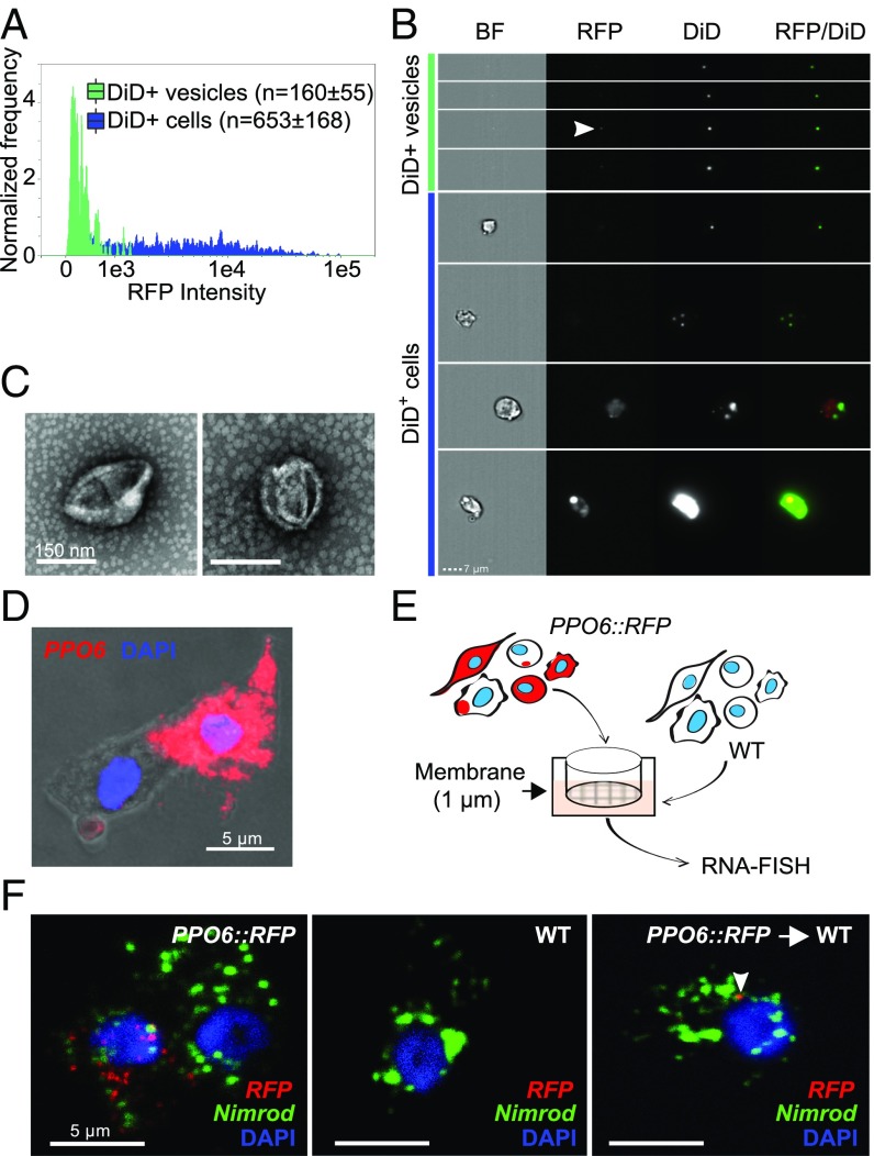
Fig. 5.
Vesicle identification and molecular exchange in mosquito blood cells. Hemocytes were perfused from PPO6::RFP mosquitoes, stained with the lipophilic DiD dye, and analyzed using imaging flow cytometry. DiD-positive cells and EVs were first identified according to their DiD and dark-field intensity. EVs were further separated from cells based on their small bright-field area. Cells were identified considering their area and aspect ratio. An RFP fluorescence histogram for both DiD-positive cells and EVs is shown in A. (B) Representative images of DiD-positive cells and EVs. The arrowhead indicates a representative of an RFP-positive vesicle. (C) Negative staining electron microscopy of vesicles obtained by differential centrifugation (10,000 × g) of hemolymph perfusate (representative images of two independent experiments are shown). (D) PPO6 mRNA detection by RNA-FISH within budding extensions of blood cells. (E) Schematics of a transwell assay developed to test the transfer of RFP between blood cells from PPO6::RFP transgenic mosquitoes and WT mosquitoes that do not express any reporter genes. Hemolymph perfusate from WT mosquitoes was pipetted onto a coverslip placed under a 1-µm membrane. Perfusate collected from PPO6::RFP mosquitoes was placed on top of the membrane. (F) RNA-FISH was performed on coverslips obtained from the transwell assay described in E using probes to detect RFP and Nimrod expression in WT acceptor cells (Right, arrowhead). Representative images of two independent experiments are shown. (Magnification: B, 63×.)

