Fig. 3.
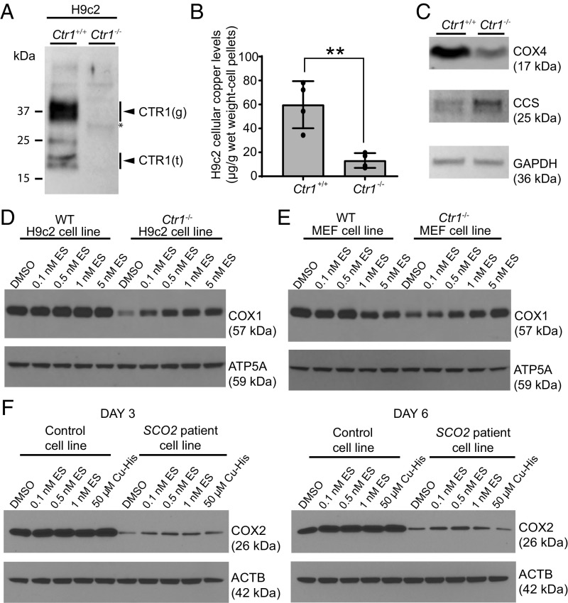
ES supplementation rescues the steady-state levels of copper-containing subunits of CcO in mammalian cell lines with genetic defects in copper metabolism. (A) Immunoblot analysis of CTR1 in Ctr1+/+ and Ctr1−/− H9c2 cells. The arrowheads labeled “g” and “t” indicate the full-length glycosylated and truncated forms of CTR1, respectively. (B) Total copper levels measured by ICP-MS in H9c2 cells. Data are presented as mean ± SD (n = 4, two-tailed unpaired Student’s t test, **P < 0.001). (C) Immunoblot analysis of CCS, COX4, and GAPDH protein levels in Ctr1+/+ and Ctr1−/− H9c2 cells. GAPDH serves as a loading control. (D) The Ctr1+/+ and Ctr1−/− H9c2 rat cardiomyocytes and (E) MEFs were cultured for 3 d with the indicated doses of ES followed by Western analysis of COX1 protein levels. ATP5A is used as loading control. (F) Control (MCH46) and SCO2 patient cell lines were cultured for 3 or 6 d in the presence of the indicated concentrations of ES or a copper–histidinate complex (Cu-His) in DMEM with 10% FBS. The cellular COX2 levels were detected by SDS/PAGE/Western blot analysis. β-Actin (ACTB) was used as a loading control.

