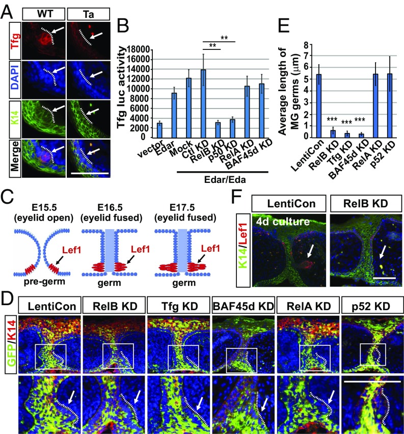
Fig. 4.
Eda-induced Tfg recruits the BAF complex and promotes MG growth. (A) IHC shows the staining of Tfg (red) and K14 (green) in WT eyelids at E15.5. (B) Luciferase activity of the Tfg promoter in Kera308 cells with indicated treatment. Data are mean ± SEM for triplicate samples. **P ≤ 0.01, Student’s t test. (C) A schematic showing the development of an MG germ (red, Lef1+) from E15.5 (eyelid open) to E17.5 (eyelid fused). (D) IHC staining of GFP (green) and K14 (red) in cultured WT eyelids transduced by lentivirus coding a GFP marker and scrambled shRNA (LentiCon) or shRNA against each indicated protein. Enlarged views of the areas marked by white rectangles are shown below each image. (E) Average length of MG germs in D. Error bars indicate mean ± SEM from at least 15 MGs of total three cultures. ***P ≤ 0.001, Student’s t test. (F) IHC of K14 (green) and Lef1 (red) in LentiCon- or RelB KD-treated eyelids cultured for 4 d. Arrows, MG germs; dotted lines separate MG germs and dermal cells. (Scale bars, 50 μm.)

