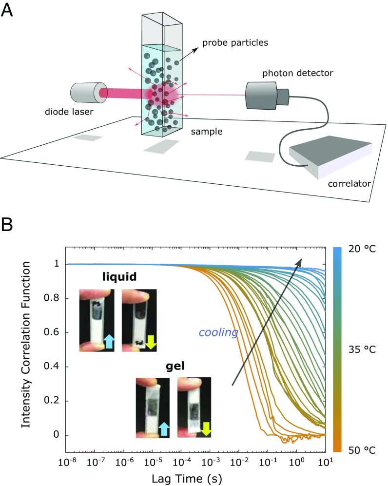
Fig. 2.
(A) Schematic illustration of the DWS setup. A 685-nm diode laser beam impinges on the sample, and the diffusely scattered light is collected by the photodiode on the other side of the sample. Tracer particles are uniformly embedded inside the sample. (B) Temperature-dependent ICF curves measured for the 500 M DNA hydrogel containing 1 (vol/vol %) 600-nm-large sterically stabilized PS tracer particles. The ICF curves were measured starting from 50 ○C (orange lines) in 1 ○C steps, cooling down to 20 ○C (blue lines). The photographs show the sample cuvette showing the samples’ liquid state at 50 ○C and the gel state at 20 ○C.

