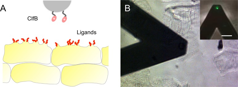
FIG 1 .
Exploring the forces between Staphylococcus aureus and AD corneocytes. (A) We used AFM-based single-cell force spectroscopy and multiparametric imaging to study the forces between bacterial cells and skin samples from AD patients immobilized on tape strips. Single bacterial probes were prepared by attaching S. aureus bacteria onto AFM cantilevers. (B) Optical microscopy image showing the bacterial AFM probe scanning across the surfaces of corneocytes. (Inset) Fluorescence image of the probe showing that the bacterial cell is alive (BacLight viability kit) (bar, 20 µm).

