Fig. 3.
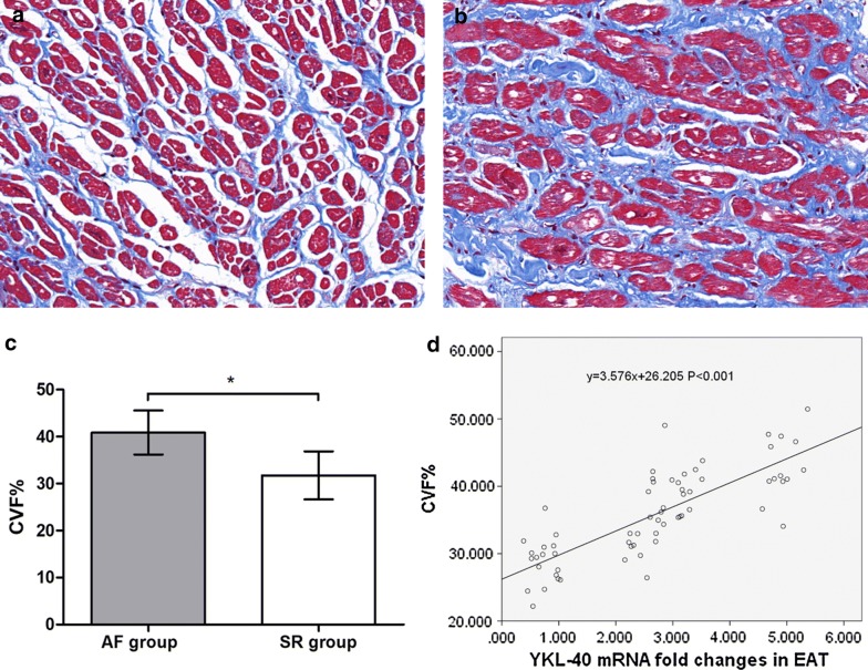
Masson staining and quantitative results of atrial myocardium from patients with atrial fibrillation (AF, n = 28) and sinus rhythm (SR, n = 36). a Masson staining of representative sections from the SR group. All cardiomyocytes were regularly arrayed with little interstitial fibers. b Masson staining of representative sections from the AF group. The cardiomyocytes were disrupted by increased interstitial fibers. c Quantitative results of Masson staining. The collagen volume fraction (CVF%) of AF group was significantly higher than SR group. *P < 0.001. d Univariate linear regression curve of YKL-40 (CHI3L1) mRNA expression (independent variable) and CVF% (dependent variable)

