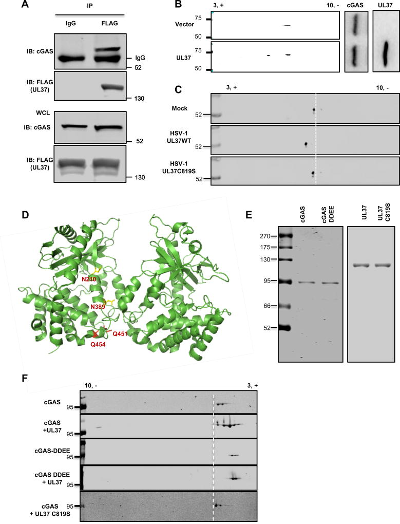
Figure 4. UL37 deamidates cGAS in cells and in vitro.
(A) Human THP-1 monocytes were infected with HSV-1 carrying FLAG-tagged UL37 at MOI=0.5. At 16 hours post-infection (hpi), cells were harvested and whole cell lysates (WCLs) were precipitated with anti-FLAG (UL37) or mouse IgG as a control. Precipitated proteins and WCLs were analyzed by immunoblotting with antibody against cGAS or FLAG (UL37).
(B) 293T cells stably expressing FLAG-tagged cGAS were transfected with a vector or that containing UL37. At 30 hours post-transfection, WCLs were prepared and analyzed by two-dimensional gel electrophoresis (2-DGE) and immunoblotting with the indicated antibodies.
(C) Primary bone marrow-derived macrophages were infected with HSV-1 (MOI=10). At 4 hpi, cells were harvested and WCLs were analyzed by 2-DGE and immunoblotting with anti-cGAS antibody.
(D) The structure of a cGAS dimer with the four deamidated residues highlighted in yellow (N210 and N389) and red (Q451 and Q454) (PDB:4K9B).
(E, F) Purified cGAS or cGAS-DDEE (as fusion constructs with maltose-binding protein from E.coli), UL37 and its deamidase-deficient UL37C819S mutant were analyzed by silver staining (E). In vitro deamidation reactions were analyzed by 2-DGE and immunoblotting (F).
Data are representative of three independent experiments (A–C, E, F).
See also Figure S5.

