
Figure 3. LGE RGs During Late Phase Lineage Progression Give Rise to Ascl1+/Dlx1+ bIPs Committed to the Generation of Matrix SPNs, See also Figures S3 – S5.
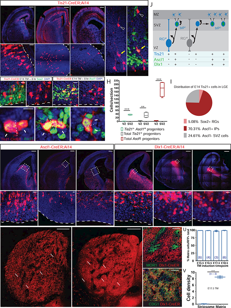
(A-C) Pulse-chase of E14.5 Tis21+ RGs and IPs at 8 (A), 24 (B), and 72 (C) hr in Tis21-CreER;Ai14 embryos. Although post-mitotic SPNs migrate into the striatum, self-renewing RGs (arrowheads) remain in the VZ for at least 72 hr (bottom panels).
(D) Example of a continually self-renewing RG in the VZ (arrowhead) 72 hr following fate-mapping from E14.5 Tis21+ RGs. Note the Ascl1+ bIP likely derived from the RG (yellow arrow) and the postmitotic progeny migrating to striatum (open arrowhead).
(E) 8-hr TM pulse-chase of Tis21+ RGs at E14.5. These RGs were Sox2+, Ascl1−, and extended end-feet to ventricle surface and radial processes (arrowheads). See also Figure S3.
(F and G) In the same sample as in (E), a large number of bIPs in the SVZ are Ascl1+ (F; yellow arrow), many of them in mitosis (G; dashed lines show plane of cytokinesis), generating putative post-mitotic progenies (arrows).
(H) Distribution of Ascl1+ IPs in VZ and SVZ following 8-hr pulse-chase in Tis21-CreER;Ai14 embryo, showing that the vast majority of Tis21+, Ascl1+ Tis21+ and Ascl1+ progenitors reside in SVZ at E14, in contrast to the distribution pattern of the same pulse-chase at E10.5 (compare with Figure 1C).
(I) Percentage of Sox2+ RGs, Ascl1+ IPs, and Ascl1- SVZ cells among the total population of fate-mapped Tis21+ progenitors at E14.
(J) A schematic showing that during the late phase of LGE neurogenesis RGs mostly generate bIPs, which may proliferate further and amplify before completing the neurogenic divisions that generate SPNs. Tis21 expression is generally activated in progenitors entering such a neurogenic phase, whether in RG or bIP.
(K-M) Pulse-chase of E14.5 Ascl1+ IPs at 8 (K), 24 (L), and 72 (M) hr in Ascl1-CreER;Ai14 embryos. Fate-mapped IPs were depleted from VZ by 24 hr after TM induction, and have differentiated into young neurons by 72 hr. Lower panels show magnified view of the boxed areas.
(N-P) 8-hr pulse-chase of Dlx1+ IPs at E13 (N), E15 (O) and E17 (P). Lower panels show that Dlx1+ IPs did not extend apical or basal processes at any of these stages and thus were exclusively bIPs. Dlx1 is also expressed in postmitotic neurons during this period.
(Q) SPNs fate-mapped from E14.5 Ascl1+ IPs localized to matrix (double arrows), but not striosomes (arrows).
(R-T) SPNs fate-mapped from E17.5 Dlx1+ IPs localized to matrix (double arrows) but not striosomes (arrows) (R); They surround MOR1+ striosomes (S) and were co-labeled by CDGI (T), with arrowheads indicating a void in the immediate region surrounding striosomes.
(U) Percentage of fate-mapped SPNs that localize to matrix following TM induction to map the output from Dlx1+ IPs at indicated embryonic times.
(V) Average cell density within the striosome versus the matrix compartment (# of cells/percent compartment area) following E17.5 TM induction in Dlx1+ IPs. ***p < 0.0001.
Scale bars: 300 µm in top of A, B, C, K, L, M, N, O, P; 20 µm in bottom of A-C, E, F, K-P; 5 µm in G; 300 µm in Q, R; 100 µm in S, T.