
Figure 12.
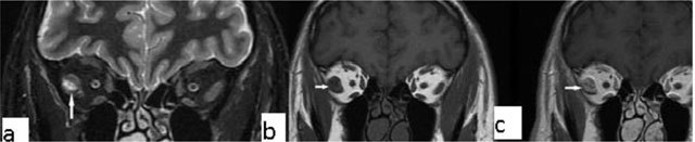
Orbital myositis. A 45-year-old female with a right orbital lesion. The lateral rectus is thickened and hyperintense on coronal fat supressed T2-WI (a, arrow). Coronal pre- and post-contrast T1-WI, showi increased enhancement of the lateral rectus (b, arrows).