
Figure 9.
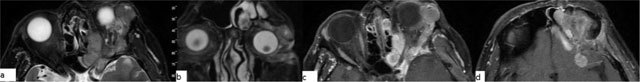
Meningioma. A 56-year-old male with a left intraconal lesion. On fat-suppressed images axial (a) and coronal (b) T2-WI, a heterogenous hyperintense mass is seen, filling the temporal fossa and orbita, extending into the frontal sinus. Post-contrast fat suppressed, axial T1-WI (c, d) show diffuse homogenous enhancement of the lesion.