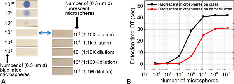
Figure 6:

Potential improvement of colorimetric assays by using fluorescent labels. (A) Blue polystyrene/latex col-orimetry microspheres and fluorescent latex microspheres spotted on nitrocellulose. 10 µL of dilutions were spotted, and the number of microspheres at each dilution is shown. (B) Detection time for fluorescent latex dilutions spotted on nitrocellulose and glass analyzed by the low-cost array platform. Our platform can reliably detect 5×10⁵ 0.5 µm microspheres on nitrocellulose membranes. This represents 2 to 3 orders of magnitude decrease in the number of microspheres detected, when compared to the number of colored latex microspheres required for a visible (positive) test line. We predict that actual assay variables could limit this number to 2 orders of magnitude.
