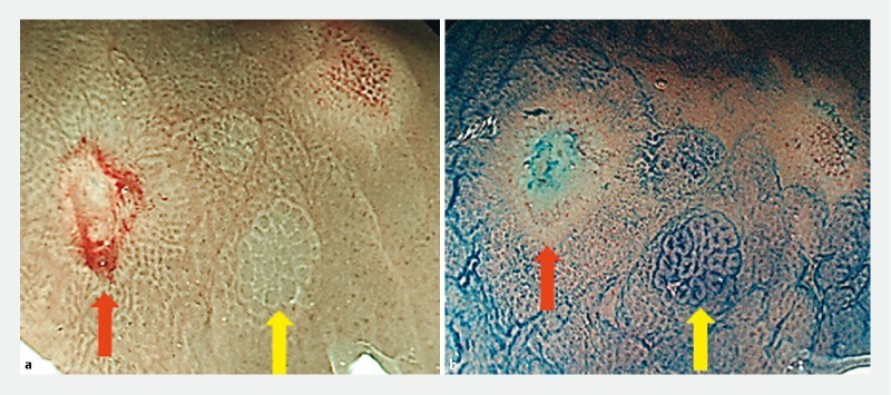
Supplementary Fig. 1 .

Identification of ACF with image-enhanced endoscopy (IEE). a ACF consisting of large crypts with white pericryptal zones were observed in the lower rectal region using magnified narrow-band imaging ( yellow arrow ). To verify the diagnosis of ACF, the adjacent mucosa was marked by argon plasma coagulation ( red arrow ). b Methylene blue staining was performed on the referral area for ACF observation. Typical ACF, which consisted of large crypts densely stained with methylene blue, were detected in the corresponding location.
