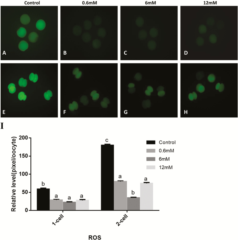
Figure 3.
(A–H) Epifluorescent photomicrographic images of in vitro matured porcine oocytes and embryos at the 2-cell stage after parthenogenetic activation. Oocytes and the 2-cell stage embryos derived from control group and various concentration glycine groups were stained with 2′,7′-dichlorodihdrofluorescein diacetate to detect intracellular levels of reactive oxygen species (ROS), respectively. (I) Fluorescence intensities were correlated with intracellular levels of ROS. Bars (a, b, and c) for adjacent pairs of columns, means without a common letter differed (P < 0.05).

