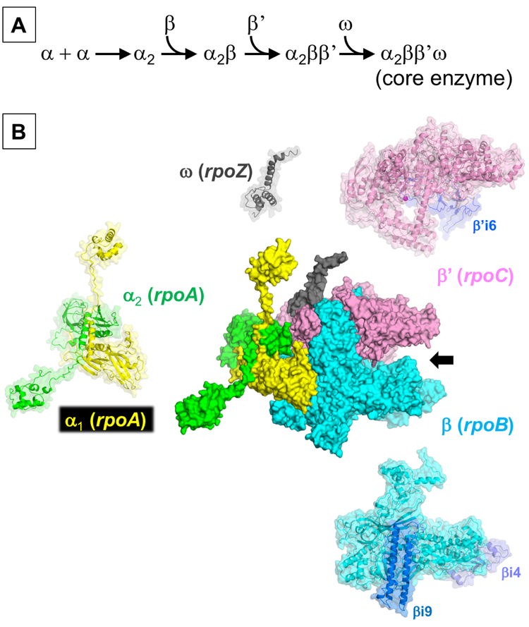
Figure 1.
A) Assembly scheme of the RNAP core enzyme. B) Structural overview of the E. coli RNAP core enzyme shown as a molecular surface representation (α1: yellow, α2: green, β: cyan, β’: pink, and ω: gray) (PDB: 4YG2). The DNA binding main channel is indicated by a black arrow. Individual subunits are also depicted with partially transparent surface to expose the ribbon model inside. Lineage specific insertions found in the β (βi4 and βi9) and β’ subunits (β’i6) are indicated in blue. The active site is represented by the catalytic Mg2+ ion (magenta sphere) coordinated in the β’ subunit.

