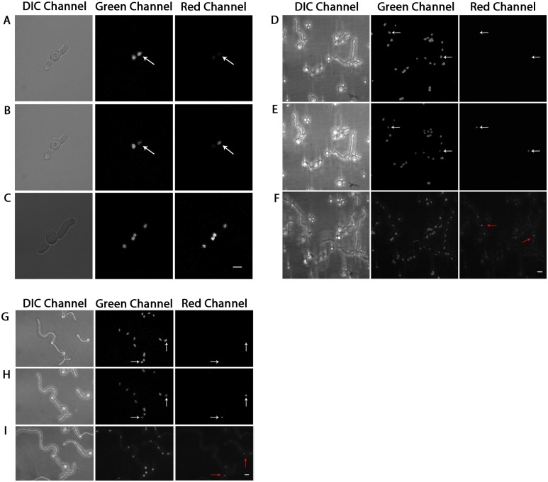
Fig 1. Histone H1 diffusion between nuclei can occur within cellular compartments.
Dendra-H1-tagged nuclei in three independent experiments, shown in DIC, green, and red channels respectively, were photoconverted using 405nm laser on an LSM-510 confocal fluorescence microscope under 100X objective lens. (A,D,G) Shows pre-photoconversion; (B,E,H) post-photoconversion; (C) post-photoconversion after approximately 3 hours have elapsed; (F) post-photoconversion after approximately 3 hours have elapsed; (I) post-photoconversion after approximately 2.75 hours have elapsed. White arrows denote nuclei which have been selected for photoactivation. Red arrows denote the location of nuclei post photoactivation. Brightness increased for entire panel images accordingly to improve visualization. White Scale Bars = 5μm.

