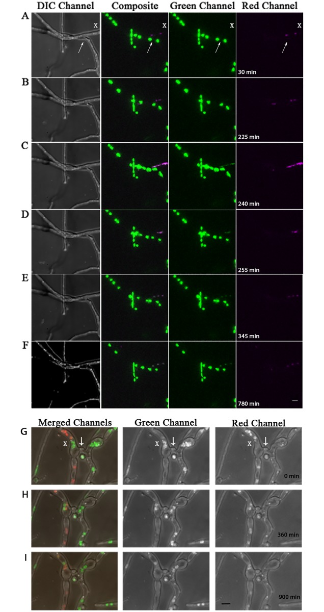
Fig 2. Histone H1 diffusion between genetically distinct nuclei can occur within cellular compartments of heterokaryotic mycelia.
Still images of DIC, merged, green and red channels from supplemental video S2 Fig, showing histone H1 diffusion within vegetative mycelia of a heterokaryon fusion between histone H1-GFP and H1-RFP-containing strains. (A-F) Showing time points: [30min, 225min, 240min, 255min, 345min and 780min] from video S2 Fig respectively (15 min/frame). Still images of merged, green, and red channels from supplemental video S1 Fig show interaction between histone H1 within vegetative mycelia of H1-GFP and H1-RFP heterokaryon fusion. (G-I) Showing time points: [0min, 360min, and 900min] from video S1 Fig (15 min /frame). White arrows denote the junction of anastomosis. White X’s mark the cellular compartments where histone H1 diffusion between genetically distinct nuclei occurs. Scale bars = 5μm.

