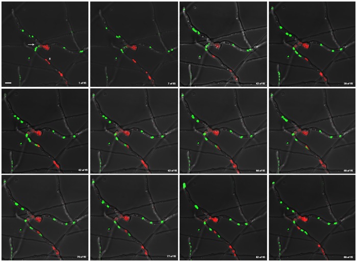
Fig 3. Genetically distinct nuclei can share cellular compartment of heterokaryotic mycelia without apparent histone H1 diffusion.
Still images of merged red and green channels from supplemental video S3 Fig, showing histone H1 diffusion within vegetative mycelia of a heterokaryon fusion between histone H1-GFP and H1-RFP-containing strains. Images show frames [1/95, 7/95, 42/95, 58/95, 62/95, 63/95, 66/95, 68/95, 70/95, 77/95, 82/95, 88/95] from video S3 Fig respectively. (Continuous imaging between frames for the duration of approximately 95 minutes). White Arrow denotes the junction of anastomosis between homokaryotic mycelia. White X marks the cellular compartment where H1-GFP and H1-RFP-containing nuclei interact. White scale bar = 5μm.

