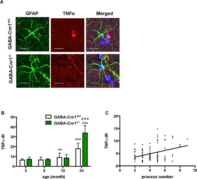
Fig 6.
(A) Representative photomicrographs of GFAP- and TNFα-immunopositive astrocytes in the stratum lacunosum moleculare layer of the CA1 hippocampal region of 24-months-old GABA-Cnr1+/+ and GABA-Cnr1−/− mice, respectively. Scale bars, 25 μm. Negative controls were stained without the anti-TNFα antibody. (B) Age-related increase in astrocytic TNFα levels is exacerbated in GABA-Cnr1−/− mice. n = 42–60 per age and genotype. +++ p < 0.001 significantly different compared to GABA-Cnr1+/+ mice from the same age group. ** p < 0.01; *** p < 0.001 significantly differs from 2-months-old mice with the same genotype. Columns represent mean values, error bars standard error of means (SEM). (C) Positive relation between the number of GFAP-positive processes and TNFα-immunoreactivity in hippocampal astrocytes of 24 months-old GABA-Cnr1-/- and GABA-Cnr1+/+ mice; n = 105.

