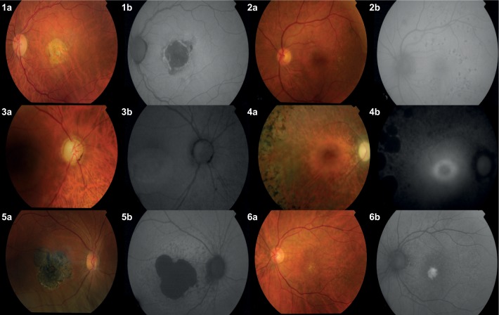
Figure 3.
Color (a) and FAF (b) imaging of different retinal conditions: 1) Exudative age-related macular degeneration, 2) diabetic retinopathy, 3) central serous chorioretinopathy, 4) retinitis pigmentosa, 5) Stargardt disease, and 6) best vitelliform disease. All images have been provided by Clínica Universidad de Navarra.
Abbreviation: FAF, fundus autofluorescence.

