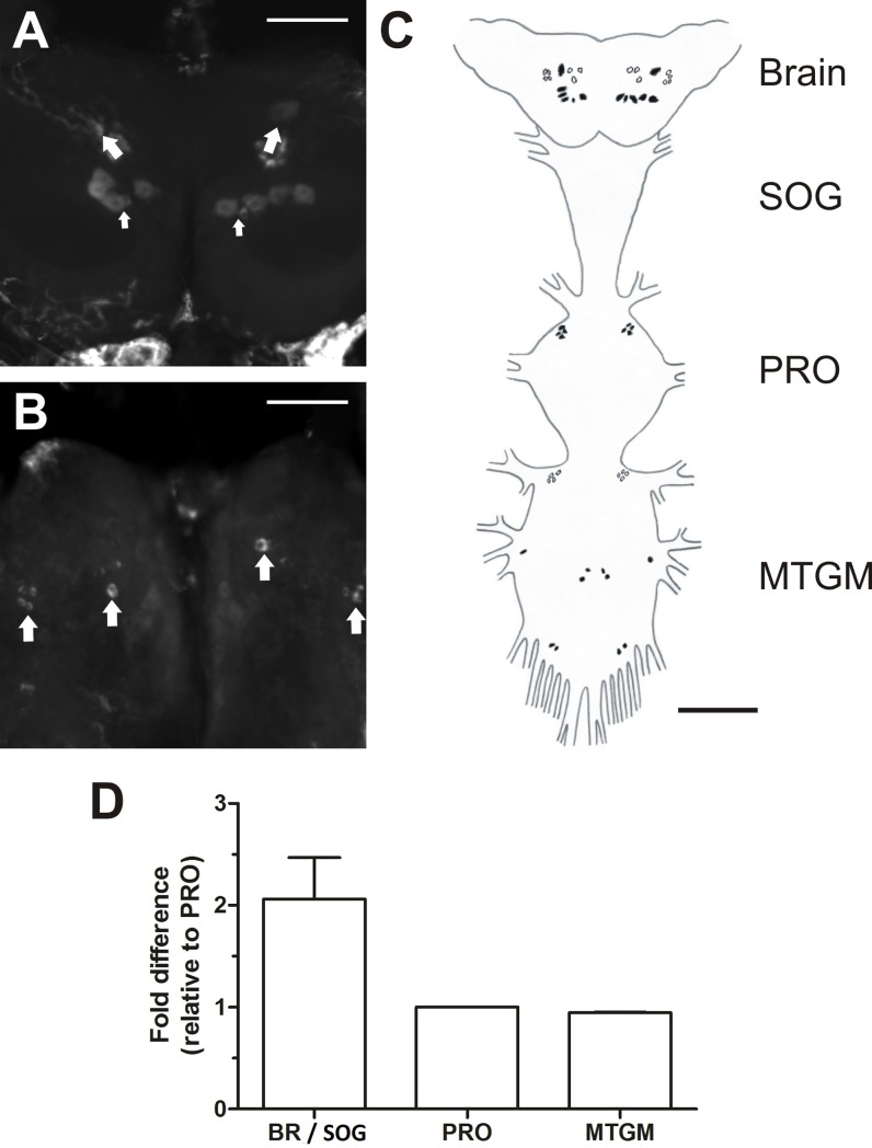
Fig 8. Cell-specific expression of the RhoprNPFR transcript in the CNS of adult R. prolixus.
(A) A stacked image showing the 6 stained dorsal bilaterally-paired neurons in the brain (indicated by larger arrows), where one paired neuron (indicated by the smaller arrows) is substantially smaller in size than the remaining 5 neuron pairs. (B) Ventral view of the brain showing clusters of 3 bilaterally-paired medial neurons and 4 bilaterally-paired lateral neurons. Each cluster is indicated by a large arrow. Scale bars represent 100 μm. (C) A schematic map of the CNS outlining the distribution of all detected neurons that exhibit RhoprNPFR transcript expression, where dorsally located neurons are represented by closed circles and ventrally located neurons by open circles. Clusters of paired cells expressing RhoprNPFR transcript are present in the PRO as well as the MTGM. Scale bar for schematic map represents 200 μm. (D) Twice as much RhoprNPFR transcript is detected in the brain and SOG compared to the PRO and MTGM. Abbreviations: BR, brain; SOG, suboesophegeal ganglion; PRO, prothoracic ganglion; MTGM, mesothoracic ganglionic mass.

