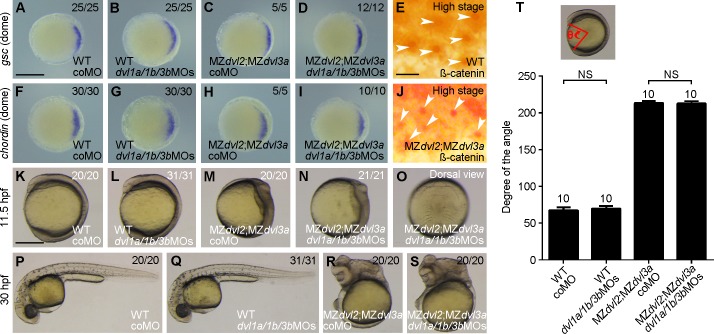
Fig 6. Knockdown of other dvl genes has no effect in MZdvl2;MZdvl3a mutants.
The embryos were injected with coMO (2.5 ng) or a mixture of equal amount of dvl1a/1b/3bMOs (6 ng in total) at 1-cell stage, except for immunostaining. All mutant embryos were imaged after different analyses, followed by genotyping. (A-D) In situ hybridization analysis of goosecoid (gsc) expression in indicated embryos at dome stage. Animal pole view with dorsal region on the right. (E) Endogenous ß-catenin nuclear accumulation (arrows) in dorsal marginal cells of a WT embryo at high stage. (F-I) In situ hybridization analysis of chordin expression in indicated embryos at dome stage. Animal pole view with dorsal region on the right. (J) Endogenous ß-catenin nuclear accumulation (arrows) in dorsal marginal cells of an MZdvl2;MZdvl3a mutant at high stage. (K-O) Phenotypes of indicated embryos at 11.5 hpf. Lateral view, with a dvl1a/1b/3bMOs-injected MZdvl2;MZdvl3a embryo also shown in dorsal view (O). (P-S) Phenotypes of indicated embryos at 30 hpf. Lateral view, note that injection of dvl1a/1b/3bMOs does not change the phenotype of WT and MZdvl2;MZdvl3a embryos. (T) Statistical analysis of the extent of axis extension delay in indicated embryos at 11.5 hpf. Bars represent the mean ± s.d. from indicated numbers of embryos (NS, not significant). Scale bars: (A-D, F-I) 400 μm; (E, J) 25 μm; (K-S) 400 μm.

