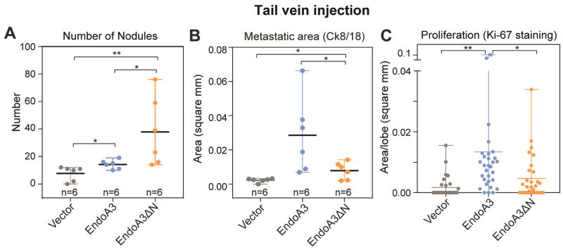
Figure 7. EndoA3 expression promotes tumor spread and proliferation at secondary sites in mice.
Experimental metastasis was examined in nude mice injected with DLD1 cells expressing vector control (gray), EndoA3-mCherry (blue), and EndoA3ΔN-mCherry (orange), respectively. (A) Number of metastatic nodules per mice in lungs upon tail vein injection of DLD1 cells. Metastatic nodules were visualized by immunofluorescence staining against Ck8/18. (B) Average area per nodule per mice in lung was measured. “n” indicates the number of animals. (C) The extent of cell proliferation was assessed using Ki-67 immunohistochemistry of pulmonary lobes. One-way ANOVA was used for multiple comparisons followed by unpaired Student's t-test was used for statistical analysis (*** p<0.001, ** p<0.01, * p<0.05).

