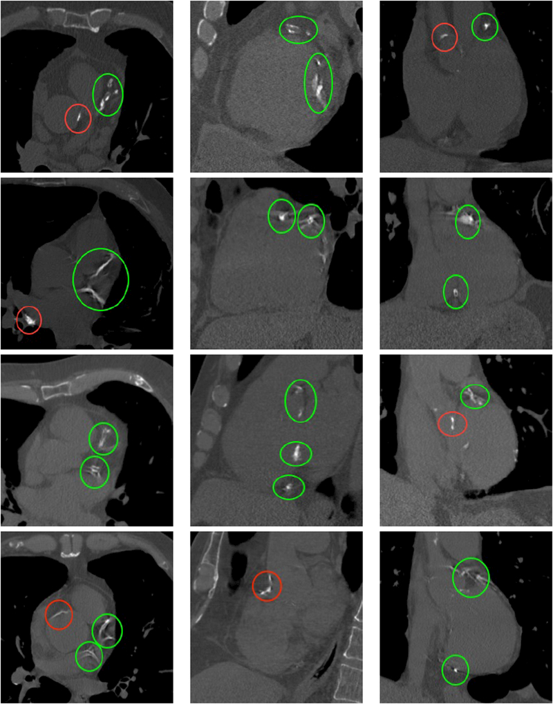
Figure 2.

Cut planes of the images of four different subjects with calcifications. First column: axial, second column: sagittal, third column: coronal. The subjects have Agatston scores of 3578, 3151, 4147 and 4217 respectively. Calcifications appear as bright structures within the coronary arteries and are highlighted with green ellipses. Please note the presence of extra calcifications in the aorta and heart valves, highlighted with red ellipses, and bone structures, such as the sternum or the vertebrae.
