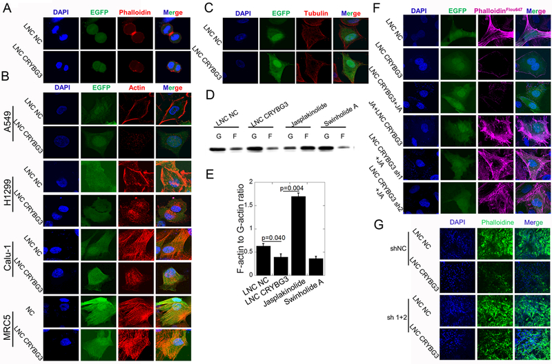
Figure 3. LNC CRYBG3 regulates cytoskeleton formation.

(A) Contractile ring dyeing in mitotic telophase. (B&C) Immunofluorescence analysis determined the microfilaments and microtubules morphology after cells overexpressed LNC CRYBG3. GFP shows the LNC CRYBG3 positive cells. (D&E) F-actin and G-actin protein levels in the A549 cells overexpressed with LNC CRYBG3 or negative control. (F) Immunofluorescence analysis determined the microfilaments morphology. (G) Frozen sections of tumors were stained with Alexa Fluor® 647 phalloidine to indicate the microfilaments and nuclear were stained with DAPI. Data represent the mean ± SE of three independent biological experiments.
