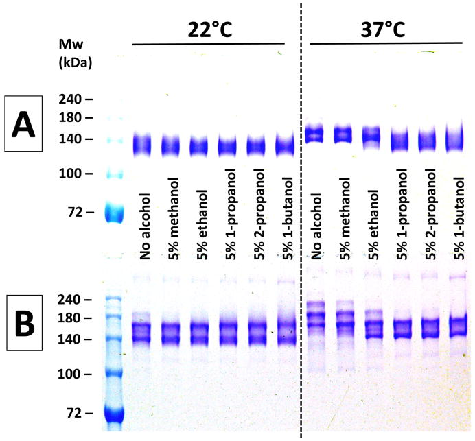
Figure 1. The effect pre-incubation with alcohols at 22°C and 37°C on non-reducing SDS-PAGE analyses of antibodies.
Panel A (top) – results using h2E2 mAb, Panel B (bottom) – results using human IgG1 polyclonal antibody. A 1.5 mm thick, 7% acrylamide gel was loaded with 2 μg (10 μL) of antibody per well, electrophoresed until the tracking dye reached the bottom of the gel, and stained with Coomassie blue. Only the top portion of the gels are shown – no bands were detected in the lower section of the gels. Panels A and B depict photographs of single gels. Pre-electrophoretic incubation in sample buffers under the indicated conditions was done for 10 minutes at either 22°C or 37°C, as indicated in the figure.

