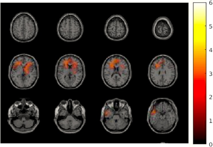
Fig. 3.
Difference of changed brain activity during resting state comparing rifaximin versus placebo. A cluster including the left inferior and middle frontal cortex, the bilateral superior frontal cortex, the right middle cingulate gyrus, and the left insula showed significantly more increased power in alpha band (11 Hz), p < 0.05

