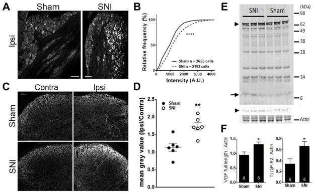
Figure 3. Spinal TLQP-62 is elevated during the establishment of neuropathic pain.
A) Representative confocal images of anti-AQEE30 immunoreactivity in ipsilateral DRG of mice 3 days post sham or SNI surgery. Scale bar: 100 μm. B) Cumulative frequency histogram of individual DRG neuron mean grey values (anti-AQEE30 immunoreactivity) identified in the L4 DRG of mice 3 days post sham (solid line, 6 mice) or SNI (dashed line, 6 mice) surgery. **** = P<0.0001, Kolmogorov-Smirnov test. C) Representative confocal images of anti-AQEE30 immunoreactivity in the contralateral and ipsilateral dorsal horn of mouse spinal cord at the level of L4 3 days post sham or SNI surgery. Scale bar: 50 μm. D) The average anti-AQEE30 immunoreactivity mean grey value ratio between the ipsilateral and contralateral L4 dorsal horn is elevated in SNI animals relative to sham controls 3 days post-surgery. ** = P<0.01, unpaired Student’s t-test. E) Representative Western blot with anti-AQEE30 shows C-terminal VGF fragments in ipsilateral spinal cord lysates from SNI and sham mice collected 3 days post-surgery. Full-length VGF is represented by a doublet of bands (arrowheads in Fig. 3E), where the lower molecular weight band is pro-VGF that is generated after the cleavage of the signal peptide. F) Densitometry analysis demonstrated that both full length VGF protein (left) and the C-terminal peptide TLQP-62 (right) are elevated in SNI animals compared to sham. Measurements for full-length VGF included both bands in the doublet and were performed using the low intensity image shown above the actin image.* = P<0.05, unpaired Student’s t-test. Data represented as mean +/− SEM.

