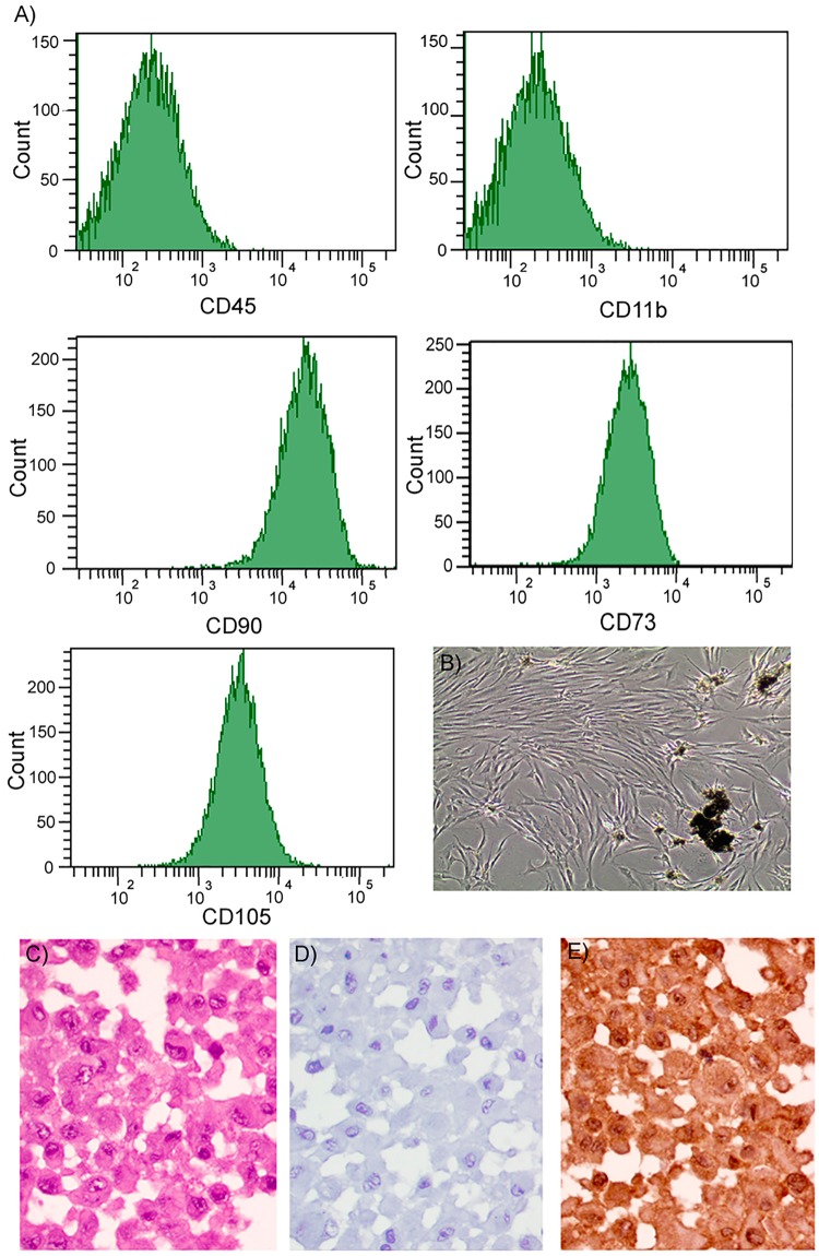
Figure 5.
(A) Flow cytometry report representing negative detection of CD11b and CD45 and positive detection of CD73, CD90 and CD105. (B) Representative image of cultured cells at initial passages with remnants of mineralized tissue. (C) H&E staining of a pellet of cultured of adherent cells showing the integrity of the cells. (D) Isotype control. (E) Nuclear and cytoplasmic detection of MSI1 (peroxidase-conjugated micropolymer) (original magnification: 40x).

