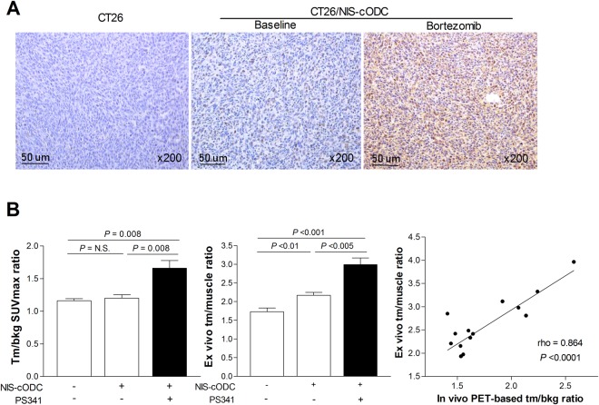
Figure 7.
Tumor NIS staining and radioiodine uptake level. (A) Immunohistochemistry for NIS expression in tumor tissues extracted after imaging. (B) In vivo tumor-to-background SUVmax ratios obtained from PET (left), and ex vivo tumor-to-muscle (tm/muscle) count ratios (middle). Bars are mean ± SD of values obtained from mice with CT26 tumors (n = 5) and mice with CT26/NIS-cODC tumors treated with vehicle (n = 6) or bortezomib (n = 8). Correlation between in vivo and ex vivo measured uptake indices with Spearman’s correlation coefficient (rho; right).

