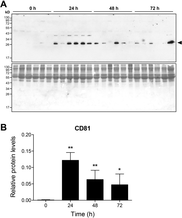
Figure 7.
Release of CD81-positive extracellular vesicles during liver regeneration. Exosomes were isolated from serum samples using ExoQuick and exosome lysates were analysed by Western blotting using an antibody against CD81 (A, upper panel). The protein band corresponding to CD81 is indicated by an arrowhead. Protein loading was controlled by coomassie blue staining of gels (A, lower panel). For each time point, samples from six individual wildtype mice are shown. Full length gels are depicted. Expression of CD81 was quantified by densitometric analyses (B). *P < 0.05, **P < 0.01 (two-tailed unpaired Student’s t-test or the Mann-Whitney U test).

