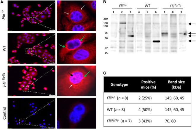
Figure 6.
Over-expression of Flii produces an altered autoantibody profile in murine OVA-induced atopic dermatitis (AD) skin-like disease. (A) Representative images of IF microscopy staining patterns produced by mouse autoreactive IgG (red) and DAPI nuclear counterstaining (blue) in primary mouse keratinocytes. Magnification 20×; scale bar 50 µm. Antibodies from OVA-induced AD skin-like lesions of Flii+/− and WT mice producing a predominantly cytoplasmic (white arrow) and perinuclear (green arrow) staining patterns, while antibodies from OVA-induced AD skin-like lesions of FliiTg/Tg mice produce a strong cytoplasmic, perinuclear (green arrow), and nuclear (black arrow) staining patterns. (B) Western blot analysis of murine keratinocyte proteins probed with pooled sera from OVA-induced AD-like mice (2–3 mice per lane); black arrows represent regions of autoreactivity. (C) Autoreactivity summary of Western blot results from murine keratinocyte lysate probed with sera from individual mice.

