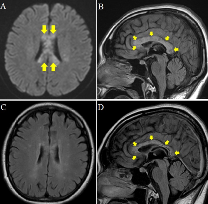
Figure 1.
Brain MRI at the beginning of hospitalization and 1 year later. (A,B) Plain MRI at the beginning of hospitalization. (A) Diffusion weighted MRI axial view showing patchy high signal intensity in the corpus callosum (arrow). (B) Fluid Attenuated Inversion Recovery (FLAIR) midline sagittal view showing edematous and irregular intensity and lower intensity at the core (arrow). (C,D) Plain MRI 1 year after hospitalization. (C) FLAIR axial view showing improved patchy high intensity in the corpus callosum. (D) FLAIR midline sagittal view shows regression of edema and ill-defined margin at the rim of splenial lesions. However, irregular signal, and low intensity at the core remained (arrow).

