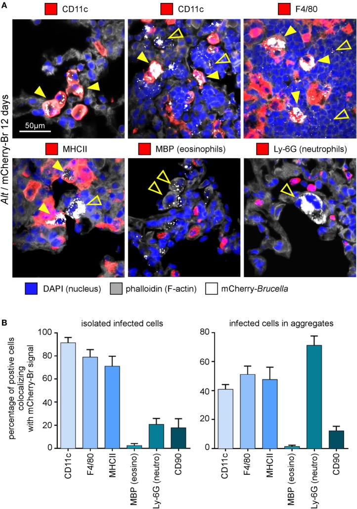
Figure 3.
Cell surface phenotype of Brucella melitensis infected cells in asthmatic lungs. Wild-type BALB/c mice received repeated i.n. administration of Alt before i.n. infection with 2 × 10⁴ CFU of mCherry-B. melitensis. The mice were sacrificed at 12 days post infection. The lungs were harvested and fixed. Frozen sections were examined by immunohistofluorescence for mCherry (Brucella), CD11c, F4/80, MHCII, major basic protein (MBP), Ly-6G, and CD90 signals. The panels are color-coded by antigen as indicated. (A) High magnification representative view of infected cells in the lungs. (B) The data represent the percentage of mCherry-Br signal that co-localizes with CD11c, F4/80, MHCII, MBP, Ly-6G, and CD90 markers. When >12 infected cells are observed in the same observation field (approximately 700 μm × 500 μm), they are considered as “aggregated,” and when <12 are observed, they are considered as “isolated.” At least 200 infected cells from three different infected mice were analyzed for each staining. These data are taken from two independent experiments.

