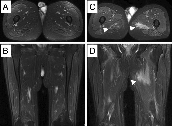
Figure 2.
Contrast-enhanced magnetic resonance images (MRI) of the lower limbs at the time of admission (A, B) did not show muscle inflammation. T1-weighted contrast-enhanced MRI axial (C) and coronal (D) images of the lower limbs on re-admission showed hyperintense signals from the internal obturator muscles to the quadriceps, bilaterally (arrowheads).

