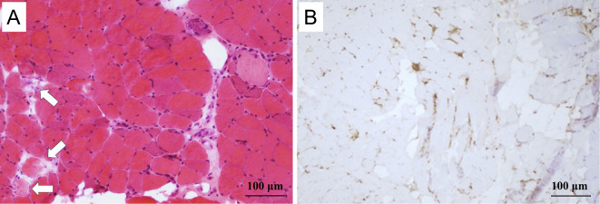
Figure 3.
Hematoxylin and Eosin staining of a biopsy of the left quadriceps muscle showed many fibers that were undergoing degeneration (arrows), as well as regenerated fibers; however, the inflammatory cell infiltration was mild (A). Many CD68+ cells (macrophages) were present at the sites of the degenerating muscle fibers (B), indicating necrotizing myopathy.

