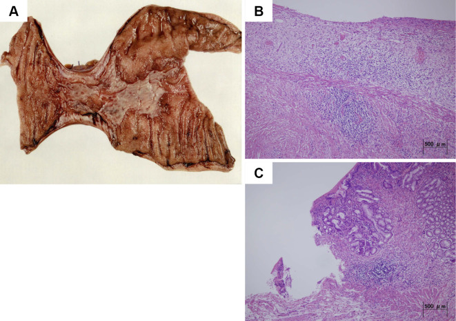
Figure 5.
Histological findings. Macroscopic examinations of the resected specimen revealed a 60-mm circumferential ulcer of grade Ul-IIIs with a partial stricture (A). Microscopic examinations of the resected specimen revealed chronic inflammation with lymphocytic and plasma cell infiltration and an edematous and congested submucosa. However, there was no evidence of sideroferous cells (B and C).

