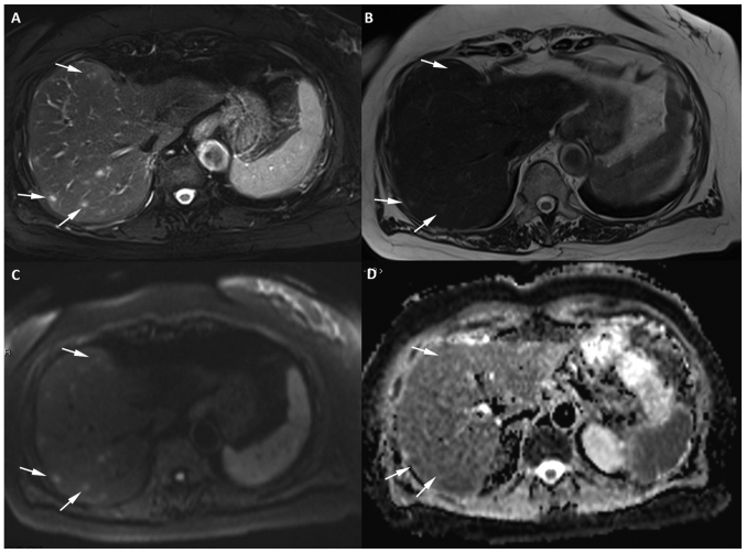
Figure 2.
Axial 1.5 T MR image of the liver with 5-mm section thickness in a patient with recurrent soft tissue sarcoma receiving treatment with Pazopanib. The image reveals a minimum of 8 focal lesions in the right lobe of the liver (three of the lesions are marked with white arrows). (A) The lesions show very high signal intensity on the T2-weighted fat-suppressed sequence and (B) mild hyperintensity on the T2-weighted sequence when compared with the background liver. (C) The lesions show high signal on DWI at b=800 smm−2 and (D) mild restriction on the apparent diffusion coefficient map. DWI, diffusion weighted imaging; MR, magnetic resonance.

