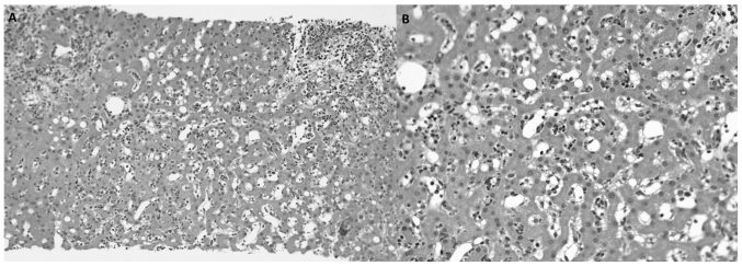
Figure 4.
Microscopic observation of the liver specimen with hematoxylin and eosin staining following the biopsy of several hepatic nodules. (A) Inflammatory portal infiltration with preservation of the limiting plate, sinusoidal dilatation and slight macrogotular steatosis are observed (magnification, ×100). (B) Lobular lymphoeosinophilic inflammatory infiltration with sinusoidal dilatation is present. Liver trabeculation is preserved. There is no evidence of malignancy (magnification, ×200).

