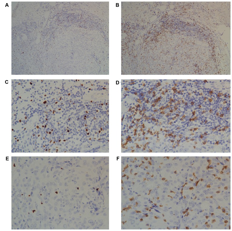
Figure 1.
Immunohistochemical analyses of intratumoral and peritumoral lymphocytes. Immunohistochemical staining of tumor-infiltrating (A) Foxp3+ T cells and (B) CD8+ T lymphocytes in invasive breast cancer. A diffuse pattern of intratumoral lymphocytes was observed. By contrast, peritumoral lymphocytes formed a lymphoid aggregate (×10 objective lens). There were markedly more Foxp3+ Tregs in the (C) peritumoral area than the (E) intratumoral areas. There was an increased number of CD8+ lymphocytes in the (D) peritumoral area than the (F) intratumoral areas. A and B, ×10 objective lens; B-E, ×40 objective lens. CD, cluster of differentiation; Foxp3, forkhead box 3.

