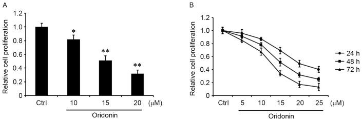
Figure 1.
Effect of oridonin on the proliferation of central neurocytoma cells. The cells were seeded in 96-well plates. Following 24 h incubation, the cells (A) were treated with 10, 15 and 20 µM oridonin or DMSO for 48 h or (B) 5, 10, 15, 20 and 25 µM oridonin or DMSO for 24, 48 and 72 h. Cell proliferation was evaluated by MTT assay and compared with DMSO-treated control cells. The assays were performed in triplicate. *P<0.05 and **P<0.01 vs. control. Data are presented as the mean ± standard error of the mean. DMSO, dimethyl sulfoxide; Ctrl, control cells.

