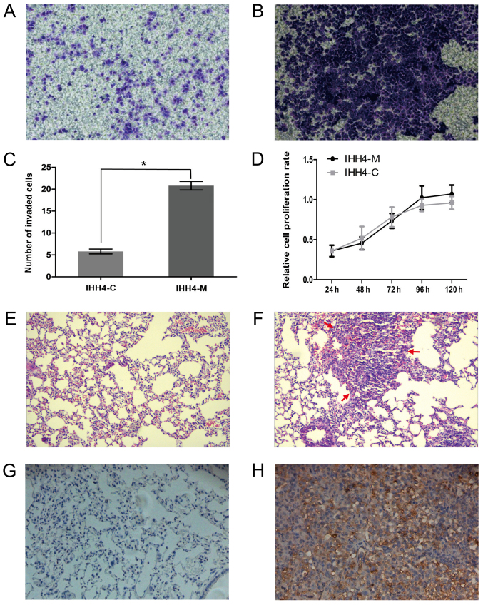
Figure 1.
The invasion and metastasis capacity of IHH4-M cells was higher than that of IHH4-C cells. The results of the Transwell assay in (A) IHH4-C and (B) IHH4-M cells (magnification, ×200). (E) No lung tumor formation was identified in the IHH4-C groups (magnification, ×100). (C) The number of cells able to invade through the Matrigel insert was significantly higher in IHH4-M cells, compared with IHH4-C cells. *P<0.001. (D) No significant difference was identified between the proliferation of IHH4-M and IHH4-C cells. (F) Apparent lung tumor formation was identified in the IHH4-M groups (magnification, ×100). CK19 protein expression was relatively low in the (G) IHH4-C groups, compared with higher levels in the (H) IHH4-M groups (magnification, ×200).

