
Figure 2.
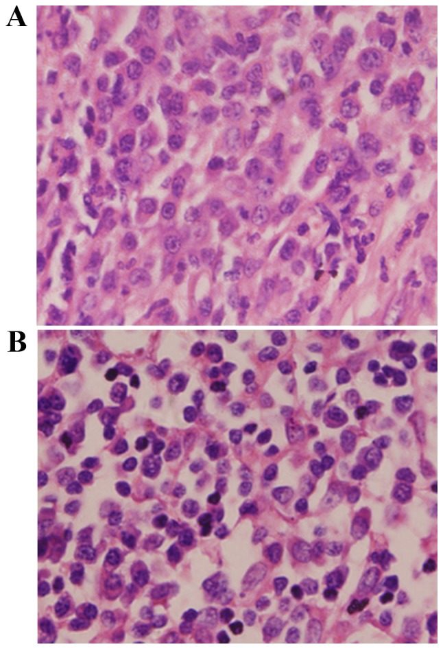
Histopathological examination of the biopsy specimens. Pathological findings revealed a large amount of plasmocyte and lymphocyte infiltration in the (A) laryngeal tumor tissue and (B) cervical lymph nodes (haemotoxylin and eosin staining, magnification, ×400).