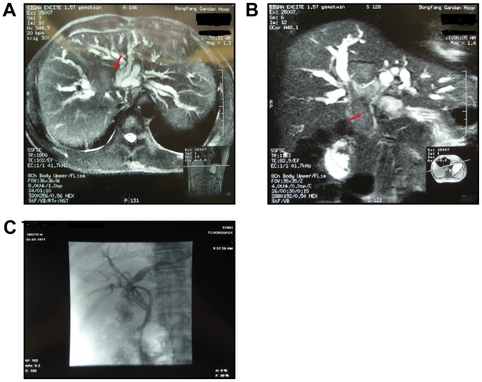
Figure 1.
Typical imaging features of hepatocellular carcinoma with bile duct invasion. (A) MRI indicates a tumor mass in the right of the liver. (B) Preoperative magnetic resonance cholangiopancreatography reveals a biliary tumor thrombus extending superficially from an intrahepatic to an extra hepatic bile duct. The red arrows indicate a biliary tumor thrombus. (C) T tube angiography was performed 2 months following surgery and revealed normal results without recurrence.

