Figure 2.
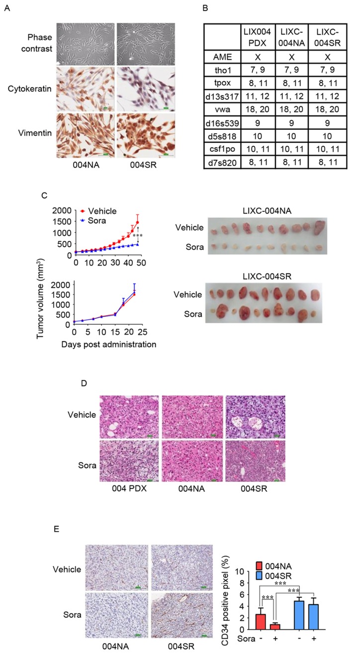
In vitro and in vivo characterization of LIXC-004NA and LIXC-004SR cell lines. (A) IHC staining of the cultured cells under phase contrast microscopy (upper panels), cytokeratin (middle panels) and vimentin (lower panels; magnification, ×200). (B) Short-tandem repeat analyses of the two cell lines and the original PDX tumor. (C) Tumor growth curves (left panels) and images (right panels) of the tumors in response to treatment with vehicle or sorafenib. (D) Hematoxylin and eosin staining of original 004 PDX tumor (left panels), 004NA tumor (middle panels) and 004SR tumor (right panels; magnification, ×100). (E) IHC using anti-CD34 antibody (left panels; magnification, ×100), and blood vessel density by quantification of the percentage of CD34+ pixels (right panel). ***P<0.001 by two-way analysis of variance. IHC, immunohistochemistry; 004 PDX, LIX-004 PDX; 004NA, LIXC-004NA; 004SR, LIXC-004SR; Sora, sorafenib; CD, cluster of differentiation.

