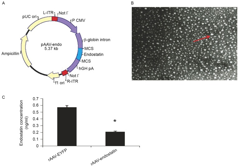
Figure 1.
Construction, production and infection of rAAV-Endostatin. (A) Construction of rAAV-endostatin vector. The endostatin gene was inserted into the MCS of pAAV vector; OS-RC-2 cells infected with rAAV-YRFP. (B) The rAAV particles in tumor cells are indicated by transmission electron microscopy (magnification, ×80,000). (C) Endostatin production in OS-RC-2 cells infected with rAAV-Endostatin was analyzed by ELISA. Comparison between RCC-rAAV-RYFP cells and OS-RC-2-rAAV-endostatin cells was performed using analysis of variance. *P<0.05. MCS, multiple cloning site; ITR, inverted terminal repeats; AAV, adenovirus-associated vector; EYFP, enhanced yellow florescent protein; ori, origin of replication.

