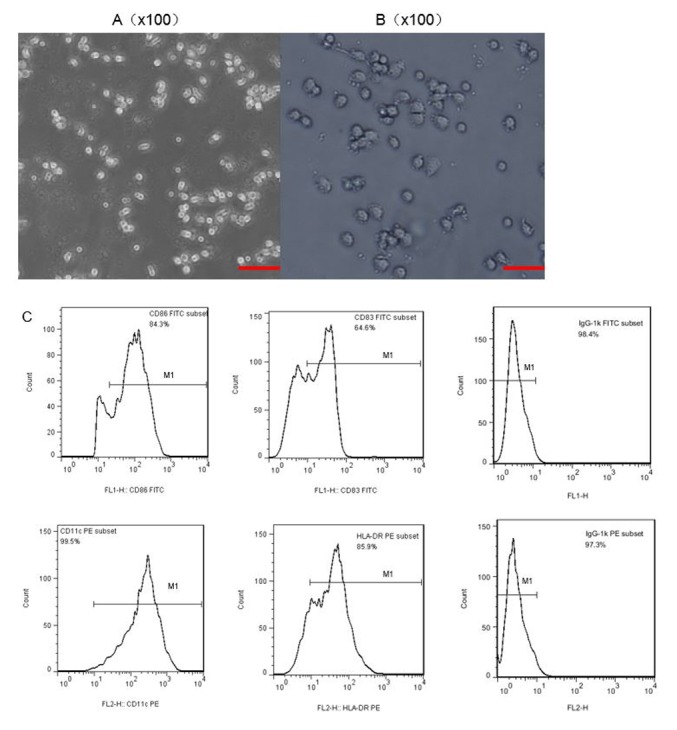
Figure 2.

Morphology and characteristics of DCs in different phases of culture. Microscopy was used to observe (A) immature DCs cultured for 3 days. (B) Subsequent to adding tumor necrosis factor (TNF-α; 20 ng/ml) on day 5 and culturing for a further two days, mature DCs could be observed on day 7. Magnification, ×100; scale bar, 100 µm. (C) Characteristic phenotypes of mature DCs were observed by flow cytometry. DCs, dendritic cells.
