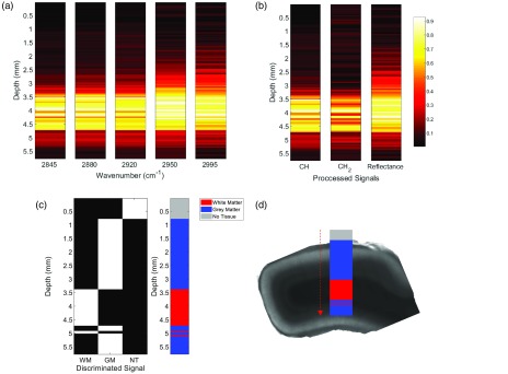
Fig. 4.
Tissue discrimination using a selection of five wavelengths along a probe trajectory through fixed primate cortex. (a) Heatmap of the intensity from each wavenumber logged when descending from air to gray matter to white matter. (b) Heatmap of processed data showing the relative increase in overall CARS signal deriving from all CH bonds, bonds specifically, and reflectance of fiber background. The maps in (a) and (b) are normalized from 0 to 1 in each graph for display purposes. (c) Thresholded processed data to discriminate different tissue subtypes based on the five wavelengths acquired. WM, white matter; GM, gray matter; and NT, no tissue sensed. (d) Overlay of the detected tissue subtypes on transmission image of primate cortex sample after removal from intact brain.

