Figure 2.
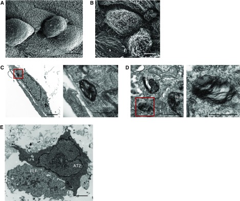
Primary AT2 cells grown in 3D organotypic coculture have preserved ultrastructural features. (A) Scanning electron microscopy of the surface of 3D organotypic coculture demonstrating the cuboidal shape of the primary AT2 cells. Scale bar: 5 μm. (B) Scanning electron microscopy of the 3D organotypic coculture showing the presence of microvilli of the AT2 cells. Scale bar: 5 μm. (C and D) Transmission electron microscopy showing the presence of lamellar bodies in AT2 cells. Panels on the right are enlargements of areas indicated by red boxes. Scale bars in C: 2 μm, with inset scale bar: 660 nm: scale bars in D: 500 nm, with inset scale bar: 1.4 μm. (E) Transmission electron microscopy shows direct contact between AT2 cells and fetal lung fibroblasts (FLF). Scale bar: 2 μm. LB = lamellar body; M = mitochondria; Nuc = nucleus.

