Figure 5.
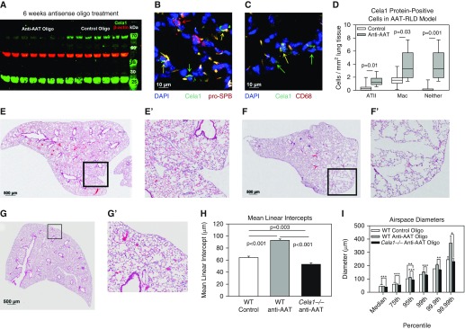
Cela1 in a murine model of AAT-deficient emphysema. (A) We previously reported a higher-than-expected molecular weight for Cela1 in mouse lung (1) and that Cela1 formed a covalent complex with AAT in vitro (2), likely accounting for this observation. A 77% reduction in this high-molecular-weight form of Cela1 was observed in anti-AAT oligonucleotide–treated lungs compared with control oligonucleotide–treated lungs (P < 0.001, Student’s t test). Little difference was noted in the quantity of native molecular weight Cela1. (B) Cela1-positive cell quantification was performed by tile scan of Cela1, prosurfactant protein B (pro-SPB), and CD68 costaining, as outlined in Figure E6. A confocal image of an anti-AAT oligonucleotide section is shown, demonstrating single-positive alveolar epithelial type II cells (ATII; red arrows), single-positive Cela1 cells (green arrow), and double-positive cells (yellow arrows). Scale bar: 10 μm. (C) Same section showing a macrophage (Mac) with Cela1 protein (yellow arrow) and nonmacrophage Cela1-positive cells (green arrows). Scale bar: 10 μm. (D) Quantification of Cela1-positive cells in control (n = 7) and anti-AAT oligonucleotide (n = 8) right lower lobe sections demonstrating increases in the number of Cela1-positive cells in the AAT-RLD model. (E) Control oligonucleotide–treated middle lobe. (F) Anti-AAT oligonucleotide–treated middle lobe. (G) Cela1−/− mice treated with anti-AAT oligonucleotide did not demonstrate any evidence of airspace injury or emphysema after 6 weeks of treatment. Scale bars: 500 μm (E, F, and G). (H) The airspaces of Cela1−/− mice treated with anti-AAT oligonucleotide (n = 7) were smaller than wild-type (WT) mice treated with either anti-AAT or control oligonucleotide. This smaller airspace size is consistent with the developmental findings outlined in Figure 1. Analysis was performed by one-way ANOVA with post hoc comparison using Dunn’s method. (I) In comparing airspace diameter percentiles, we found that the Cela1−/− anti-AAT oligonucleotide–treated mice had an airspace diameter distribution comparable to that of WT control oligonucleotide–treated mice. *P < 0.05; **P < 0.01; ***P < 0.001.

