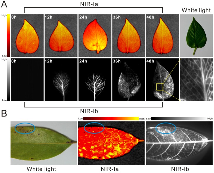
Figure 3.
Leaf vein imaging and plant anthracnose infection detection using IR-808. (A) NIR-Ia and NIR-Ib images of whole leaves picked from E. aureum at different time points post dye administration. (B) NIR-Ia and NIR-Ib imaging detected anthracnose of Khaya senegalensis A. Juss (circled in blue). The excitation wavelength was 680 nm for NIR-Ia imaging and was 808 nm for NIR-Ib imaging. A bandpass filter of 700 nm to 900 nm was used for NIR-Ia imaging and from 900-1,000 nm for NIR-Ib imaging. A fixed exposure time of 1 s was used for NIR-Ia imaging and 200 ms for NIR-Ib imaging. The power density of excitation at the imaging plane was 45 mW/cm2.

